 English
 한국어
한국어 français
 Deutsch
 Español
 русский
 português
 العربية
 ไทย
ไทย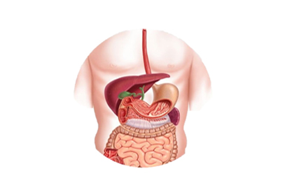
Address
1st & 2nd Floor, 10 Building, 18 Huashan Rd., Changzhou, Jiangsu province, China

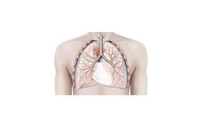
The medical endoscope is an important and commonly used medical device in clinical practice. It can enter the human body through natural cavities or small incisions produced by surgery, allowing doctors to make diagnoses and treatments under direct vision. It is widely used in departments such as general surgery, urology, gastroenterology, and respiratory medicine. With the advancement of technology, the history of medical endoscopes can be roughly divided into four stages: the rigid endoscope period, the semi-flexible endoscope period, the fiber endoscope period, and the modern endoscope period.
In the early stage of open rigid tube endoscopes, German physician Philip Bozzini first proposed the idea of an endoscope in 1804, and in 1806, he manufactured an instrument with a candlelight source that consisted of a vase-shaped light source, a candle, and a series of lenses, which was used to observe the bladder and rectum of animals. Although it was not used in humans, Bozzini is still recognized as the inventor of the first endoscope.
In 1879, Berlin urologist Max Nitze made the first endoscopy product (i.e. cystoscope) with an optical system, which contained a prism at the front end and used electricity to heat a platinum wire loop to create light. This endoscope was only used for the urinary system. Although this endoscope could obtain clearer images, it still had obvious shortcomings such as the use of lenses that could cause perforation, and the inevitable risk of burns and complications, which caused patients pain and fear. With the rapid development of technology, clinical doctors and scientists continued to explore, and the technology of this diagnostic tool was improved to a new level.
The first truly semi-flexible endoscopy product appeared in 1932. This semi-flexible gastroscopy was developed by Schindler in cooperation with a device maker starting in 1928, and was named the Wolf-Schindler gastroscopy. The front end was flexible and could enter internal structures with some bends, and it became the standard product in the field of gastroscopy for more than 20 years.
Although Schindler's gastroscopy instruments achieved great success, it still had two fatal weaknesses: firstly, incandescent lamps were a type of heat source that was very unfriendly to the human body for examination; secondly, although semi-flexible endoscopes were better than rigid ones in terms of experience, they were still limited in terms of performance.
It wasn't until 1952 that a historic cold light appeared in the development of endoscopes. French scientists manufactured a glass fiber illuminator for cold light sources, which improved the safety of laparoscopy by eliminating the danger of burns and electrical malfunction caused by internal lighting sources, laying the foundation for the subsequent application of flexible fiber instruments.
In 1967, a Japanese company first used an external fiber-optic beam coupled to a strong cold light source to fundamentally improve the lighting of endoscopy products. The fiber-optic of the fiber endoscope is made from thousands of sintered glass fibers with total reflection characteristics, combining the functions of light guidance and imaging. The use of a light guide glass fiber reduced the loss of light propagation to the greatest extent possible, improving the brightness and clarity of the visual field. The extremely fine and soft light guide fibers are about 1/10 of a hair, making the endoscope slender and flexible, greatly reducing patient pain and observation blind spots. In addition, the use of an externally connected cold light source for "cold processing" technology has significantly increased the brightness of the viewing field. In summary, the use of glass fibers and cold light sources is a hallmark of fiber endoscopes, and this revolution has greatly improved the flexibility of the device, the brightness of the visual field, and the clarity of the image, making it a significant improvement in the history of endoscope development.
In 1983, the first electronic endoscope with a miniature image sensor replacing fiber-optic image bundles was developed by an American company. Optical and electrical signal conversion is performed using a photoelectric coupler device, and the image is displayed on a monitor. It can also be equipped with auxiliary devices for information input and diagnostic processing tools etc.
Electronic endoscopy products have high resolution, clear images, vivid colors, high intensity, long life, and are more durable. It also has features that cannot be replaced by other forms of endoscopes, including multiple person viewing and image data storage, marking a new chapter in the history of endoscope diagnosis and treatment, and playing a significant role in clinical, educational and scientific research fields.