 English
 한국어
한국어 français
 Deutsch
 Español
 русский
 português
 العربية
 ไทย
ไทย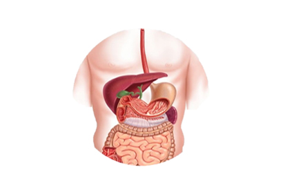
Address
1st & 2nd Floor, 10 Building, 18 Huashan Rd., Changzhou, Jiangsu province, China

(1) Polypectomy
That is, submucosal injection-resection method, separate the submucosa, inject liquid, and fully lift the lesion during full submucosal injection. If the lesion is less than 2cm, use a snare to circle it, lift it along the wall, loosen the mucosal muscle layer that may be clamped up, and then remove the lesion with EMR equipment.
(2) Transparent cap method
Transparent plastic caps with different specifications, flat or angled surfaces are installed at the inner lens end to attract and remove the lesions.
After submucosal injection of the lesion under endoscopy, put the endoscopic snare in the groove at the front end of the transparent cap. The transparent cap EMR equipment is aligned with the lesion to be removed and sleeved into the transparent cap. Tighten the snare to cut the lesion. Before electric cutting, loosen the snare slightly to restore the muscularis propria that may be involved to its original position.
(3) The ligation device method
The inner lens is equipped with a ligation device. After aiming at the lesion to be removed and attracting, the rubber band covers the lesion to form a pedicle like polyp, and then the lesion including the rubber band is electrically cut under the rubber band.
This method has the advantages of simple operation, clear visual field, easy to grasp the depth of resection, less local injury, less bleeding and complications, and is relatively safe.
(1) Mark, use a needle incision or argon ion coagulation to mark the resection area with 0.5cm electrocoagulation at the edge of the lesion;
(2) Before submucosal injection of liquid, the clinically available liquids for submucosal injection include physiological saline, glycerol fructose, sodium hyaluronate and so on.
(3) Pre-cut the surrounding mucosa: use the ESD equipment to cut part of the mucosa around the lesion along the marking point or the outer edge of the marking point, and then use the IT knife to cut all the surrounding mucosa;
(4) According to different parts of the lesion and the operation habits of operators, ESD equipment IT, Flex or HOOK knife and other stripping instruments were selected to peel the lesion along the submucosa;
(5) For wound treatment, argon ion coagulation was used to electrocoagulate all visible small blood vessels in the wound to prevent postoperative bleeding. If necessary, hemostatic clamps were used to clamp the blood vessels.
EMR and ESD derived from the same origin and have similar technical characteristics. EMR ESD difference as follows:
The disadvantage of EMR is that it is limited by the size of resectable lesions under endoscopy (less than 2cm). If the lesions are greater than 2cm, it needs to be resected in blocks, the edge treatment of resected tissues is incomplete, and the postoperative pathology is inaccurate.
However, the ESD equipment expands the indications of endoscopic resection. For lesions larger than 2cm, it can also be completely removed. It has become an effective means for the treatment of early gastrointestinal cancer and precancerous lesions.
At present, EMR and ESD are widely used in the resection and treatment of digestive endoscopy.
EMR and ESD technology is the killer of endoscopic resection, and has become an important means of minimally invasive treatment of early gastrointestinal cancer and precancerous lesions. It is believed that EMR and ESD equipment and EMR and ESD endoscopy can create greater medical value for the health of people in the future.