 English
 한국어
한국어 français
 Deutsch
 Español
 русский
 português
 العربية
 ไทย
ไทย
Address
1st & 2nd Floor, 10 Building, 18 Huashan Rd., Changzhou, Jiangsu province, China


【Case data】
A 62-year-old female, was admitted to the hospital for " right upper abdominal pain for 2 months". Complete blood biochemical examination indicated that the liver function was in the normal range, and no abnormality was found in the detection of tumor markers. MRI examination showed that nodules in the common hepatic duct were considered to be tumorous lesions, and the wall adjacent to the right hepatic duct was slightly thickened and the intrahepatic bile duct was slightly dilated. ERCP+SpyGlass +Spybite biopsy were performed to further clarify the diagnosis and the nature of the lesions.

Fig.1-2 :MRI showed dilation of the intrahepatic bile duct and nodules in the common hepatic duct. Considering the possibility of neoplastic lesions, the bile duct wall adjacent to the opening of the right hepatic duct was locally thickened.

Fig.3-4 ERCP equipment showed irregular hilar bile duct stricture, and SpyGlass was performed.
【SpyGlass Image 】:

SpyGlass shows irregularity of the hilar bile duct, and intraluminal mass with cobweb-like angiogenesis can be seen(Fig.5-6)。
A total of 8 Spybite biopsies were performed at the tumor part, the lower bile duct margin, and the tumor upper bile duct margin.(Fig.7-8)。
Hilar Tumor Biopsy Video
【SpyGlass Biopsy pathology】
Papillary adenoma, focal high-grade intraepithelial neoplasia, suspicious for cancer under endoscopy(Fig.9- 10)。
【Surgical analysis】Hepatic lobectomy (left hemihepatic and caudal lobectomy), cholecystectomy, choledochectomy, cholangioplasty, hepatobiliary-jejunostomy, portal vein exploration, and abdominal lymph node dissection were performed.
【Postoperative pathology】
.png "图片5_(1).png")
Postoperative pathology showed: 1. (Hilar) moderately differentiated villous-tubular cholangiocarcinoma, which invaded the fibrous tissue outside the muscularis layer of the bile duct. No cancerous involvement was found in any of the resection margins. There were 2 palpable lymph nodes around the common bile duct, no metastatic cancer (0/2) was found under the endoscope, and the immunohistochemical results showed: CK-P (-). 8 groups of lymph nodes (0/9) and 12 groups of lymph nodes (0/2) were sent for examination, and no metastatic cancer was found under the endoscope(Fig.11-12)。
【Discussion】
In this case, preoperative MRI showed that the hilar bile duct was occupying space in the lumen of the hepatic duct, and the tumor was likely, suggesting that the wall of the right hepatic duct orifice was thickened. Before the operation, not only the bile duct tumor, but also the right hepatic duct invasion should be identified, which is more accurate. The resection method and scope of resection were formulated according to the requirements of SpyGlass image diagnosis system and Spybite biopsy, which not only diagnosed bile duct tumors before operation, but also further diagnosed as Bismuth type II hilar cholangiocarcinoma. R0 cut off. At present, there are large and prospective series of studies, which believe that POCS is an effective and safe intervention method to guide the treatment of patients with unknown bile duct strictures [1]. However, future randomized trials are still needed to determine the optimal number of biopsies [2]
References:
1. Almadi, M.A., et al., Using single-operator cholangioscopy for endoscopic evaluation of indeterminate biliary strictures: results from a large multinational registry. Endoscopy, 2020. 52(7): p. 574-582
2. Wen, L.J., et al., Efficacy and Safety of Digital Single-Operator Cholangioscopy in the Diagnosis of Indeterminate Biliary Strictures by Targeted Biopsies: A Systematic Review and Meta-Analysis. Diagnostics (Basel, Switzerland), 2020. 10(9).
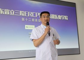
Expert introduction:

Wang Wei
Wuhan No.1 Hospital
Head of Endoscopy Group of Hepatobiliary Surgery, Wuhan No.1 Hospital
Visiting Scholar, EMORY University Hospital, USA
Member of the Perioperative Branch of the Chinese Association of Integrative Medicine
2018 National Youth ERCP Competition Youth Judges
Hubei Province ERCP Elite Member
Member of Wuhan Digestive Endoscopy Association
Member of Wuhan General Surgery Quality Control Center
