 English
 한국어
한국어 français
 Deutsch
 Español
 русский
 português
 العربية
 ไทย
ไทย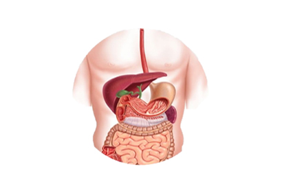
Address
1st & 2nd Floor, 10 Building, 18 Huashan Rd., Changzhou, Jiangsu province, China

The clinical manifestations of biliary and pancreatic diseases are mainly abdominal pain, jaundice and fever. A clear preoperative diagnosis is crucial to choosing a reasonable treatment plan. In recent years, with the continuous development and progress of various non-invasive imaging methods, the diagnostic level of biliopancreatic diseases has been continuously improved.
B-ultrasound is the first choice for diagnosing biliary tract diseases. It has the advantages of convenience, non-invasiveness and low cost.
However, B-ultrasound for common bile duct lesions, especially the lower segment of the common bile duct, is often missed due to duodenal gas covering and other effects, and is also affected by the operator's experience.
CT has a high resolution for solid organ lesions, but has a limited diagnostic rate for luminal lesions such as the intestine and bile duct, especially when there is no calcification in the stone.
Due to the limitations of B-ultrasound and CT, there are still some patients with obstructive jaundice. The ERCP instrument is under the X-ray monitoring, and the contrast agent is directly injected retrogradely into the cholangiopancreatography through the endoscope and the catheter.
Its image is clear, high resolution, continuous, dynamic, direct vision, not easily disturbed by intestinal gas, etc. Its diagnostic rate for bile duct stones can reach 92% to 94%, especially for some small stones.
ERCP examination can clearly indicate that there is a small filling defect in the extrahepatic bile duct. During ERCP examination, the gas must be exhausted first and the contrast medium should be injected slowly. ERCP not only has diagnostic value, but also has its therapeutic effect.
The incidence of tumors of the biliopancreatic system tends to increase, because the lower segment of the common bile duct is located in the dorsal side of the duodenum, and the deep pancreas retroperitoneum and ampulla have a special adjacent relationship with the pancreas.
In clinical practice, B-ultrasound and CT cannot provide a more accurate diagnosis for the above-mentioned space-occupying lesions, especially in cases with jaundice or intrahepatic and extrahepatic jaundice expansion, which are difficult to differentiate from common bile duct stones clinically.
The images provided by the ERCP examination can clearly and intuitively reflect the images of the bile and pancreatic ducts. The ERCP instrument is more accurate than B ultrasound in diagnosing pancreatic cancer, and the accuracy of CT can reach 95%. The bile ducts of pancreatic cancer are infiltrated by pancreatic tumors, compressed and deformed.
The stenosis of the distal bile duct (pancreatic segment bile duct) can be displayed at the same time as the pancreatic duct is visualized, which is called the "double duct sign", which has specific diagnostic significance for pancreatic cancer.
At the same time, due to the compression of the bile duct, the bile duct is passively dilated, the bile duct wall is thinned, and the bile duct is tortuous and elongated. It is called the "soft vine sign" in imaging, and it is also an ERCP sign of great significance for the diagnosis of pancreatic cancer.
Cholangiocarcinoma, due to its special anatomical location in the hilar part and the well-differentiated sclerosing cholangiocarcinoma pathological features, tends to infiltrate the hepatic bile duct or the distal bile duct along the submucosal bile duct, so it is difficult for both B-ultrasound and CT. Prompt a hilar solid mass.
The medical ERCP can completely display the entire bile duct system, and can find the filling defect of the hilar bile duct, which provides a good help for the diagnosis.